期刊名称:MOLECULAR VISION
|
ISSN: | 1090-0535
|
|
版本: | SCI-CDE
|
|
出版频率: | Continuous publication
|
|
出版社: | MOLECULAR VISION, C/O JEFF BOATRIGHT, LAB B, 5500 EMORY EYE CENTER, 1327 CLIFTON RD, N E, ATLANTA, USA, GA, 30322
|
|
出版社网址: | http://www.molvis.org/
|
|
期刊网址: | http://www.molvis.org/molvis/
|
|
影响因子: | 2.367 |
| 主题范畴: | BIOCHEMISTRY & MOLECULAR BIOLOGY; OPHTHALMOLOGY |
 期刊简介(About the journal)
投稿须知(Instructions to Authors)
编辑部信息(Editorial Board) 期刊简介(About the journal)
投稿须知(Instructions to Authors)
编辑部信息(Editorial Board)
About the journal

Molecular Vision is a peer-reviewed journal dedicated to the dissemination of research results in molecular biology, cell biology, and the genetics of the visual system (ocular and cortical). Molecular Vision is indexed in Medline, PubMed, and Index Medicus by the National Library of Medicine (NLM); in Science Citation Index, Current Contents, and other indexes by the Institute for Scientific Information (ISI); in Biological Abstracts by BIOSIS; and in Chemical Abstracts by the Chemical Abstracts Service. Access to the journal (articles and services) is free of charge.
Instructions to Authors
A major benefit of submitting a manuscript to Molecular Vision is the potential for rapid publication. For this to happen, the following instructions must be carefully followed. Departures from these instructions create more work for the editorial staff and will result in delays. Substantial departures will result in the manuscript being returned to the authors without consideration.
- Synopsis of Guidelines
- Manuscript Organization and Styles
- Preparation of Manuscripts
- Submission of Manuscripts (includes graphics considerations)
- The Review and Publishing Process
For questions not covered in these instructions, please email the Editors.
Manuscript organization and styles:
Molecular Vision encourages the submission of manuscripts on the molecular biology, the cell biology, or the genetics of the visual system (ocular and cortical). Manuscripts should present original, unpublished material not being considered for publication elsewhere and written to be accessible to vision scientists. If accepted, the material (data and text) shall not be published elsewhere without the consent of the Editors and Publisher of Molecular Vision. While there is no limit on the length of a manuscript, manuscripts are expected to be concise.
Following are descriptions of the types of manuscripts Molecular Vision accepts with an outline of the required elements for each manuscript type. Detailed descriptions of the elements of a manuscript are given in the section on "Preparation of manuscripts."
Research Article:
A detailed description of original, unpublished work covering a positive or negative result of significance. Reports of negative results must be investigations that other investigators would be likely to pursue in the absence of the report.
Molecular Vision does not publish simple reports of sequence data. Reports of new sequence (nucleotide or protein) may accompany related biological data. Simple sequencing data should be submitted to GenBank.
A research article must include the following sections: Title Page, Structured Abstract, Introduction, Methods, Results, Discussion, and References. Acknowledgements, Figures, and Tables should be included as appropriate.
Technical Brief:
This is a short account detailing a novel method or unique use of current technology that of itself, regardless of the experimental question studied, represents a significant addition to scientific enquiry.
A technical brief must include the following sections: Title Page, Descriptive Abstract, Introduction, Methods, Results, Discussion, and References. Acknowledgements, Figures, and Tables should be included as appropriate.
Review:
The Editors solicit suggestions for comprehensive articles reviewing the current status of a particular field or topic. The Editors should be contacted in advance with topics so that appropriateness for publication is confirmed. Appropriateness will be determined by the importance of the topic, lack of existing reviews on topic, and timeliness of request. Reviews of appropriate topics are still subject to peer review; a determination that a topic is acceptable does not guarantee acceptance of a manuscript.
A review must include the following sections: Title Page, Descriptive Abstract, Introduction, Discussion, and References. If Results are to be presented, the manuscript should include both a Methods and a Results section. Acknowledgements, Figures, and Tables should be included as appropriate.
Preparation of manuscripts:
The sections of the manuscript (Abstract, Introduction, Methods, etc.) should be clearly labelled with the name of the section on a line by itself. Figure legends and Tables should follow the References section. Each paragraph should be preceded and followed by a blank line. The Editors recommend that authors read the Molecular Vision Style Guide before preparing a manuscript for submission.
If any figures, graphs, tables, or data previously published are used, written permission of the publisher of the previous work must be presented. This includes works by the authors of the current submission. Obtaining this permission is the sole responsibility of the author.
Manuscript information
All manuscripts should begin with the title of the manuscript, the names of the authors, the authors' institutional affiliations, and contact information for the corresponding author. Each author may also include an email address or a web address (URL).
Abstract
The abstract should concisely (in fewer than 4000 characters) summarize the work presented. Manuscripts submitted as a Research Article must have a structured abstract consisting of four subsections: Purpose, Methods, Results, and Conclusions. Other types of manuscripts must include a descriptive abstract that details the topics covered in the manuscript.
Introduction
A succinct introduction without subheadings should describe the purpose or goals that led to the production of the manuscript. This should include a concise review of relevant literature.
Methods
This section should detail everything that would be required to replicate the work presented. Non-proprietary names should be used whenever possible. Where relevant, the name of the supplier of items used in the investigation should be identified. Suppliers should be identified by their full company name and location (city, state/country); if the company maintains an online presence, that may also be given.
Studies involving animals should include a statement that animal care guidelines comparable those published by the Institute for Laboratory Animal Research (Guide for the Care and Use of Laboratory Animals) or the US Public Health Service (Public Health Service Policy on Humane Care and Use of Laboratory Animals) were followed. Studies that involve humans subjects should indicate that an appropriate institutional review board has approved the project. If such a review process is unavailable, the authors should follow the principles of the Declaration of Helsinki (JAMA 1997; 277:925-926).
Results
The findings of the investigation should be presented without interpretation or discussion. In short manuscripts where the interpretation of the data is relatively simple, the data may be discussed in a combined Results and Discussion section.
Discussion
An interpretation and commentary on the data (research article) or technique (technical brief) should be presented without speculating beyond the scope of the investigation. For reviews, the discussion is the bulk of the manuscript. It may be sectioned to fit the topic being reviewed and each section may be titled by the author.
Acknowledgements
Authors may briefly mention individuals making significant non-authorship contributions to the manuscript. Funding support for the work presented should be detailed. Authors should also disclose any commercial interest in the subject of the manuscript or in entities discussed in the manuscript. All prior presentation of the manuscript's data at meetings should be indicated; such presentations should not appear among the manuscripts references.
References
Citations in the text are noted in appropriate places by numbers in brackets (e.g., [3,5,8-12]). References must be numbered in the order of appearance in the text of the manuscript. All references cited in the text should be listed in the References with corresponding numbers. References should follow the Vancouver style as described by the International Committee of Medical Journal Editors (Ann Intern Med 1997; 126:36-47) except for part 33 which is outdated (see Example 4 below). This style orders elements of the source of journal articles from least to most specific and has been adopted by the National Library of Medicine.
References to unpublished work should be made parenthetically in the body of the text and not listed as a citation in the References section. If the data comes from some or all of the authors of this work, you may simply list it as "[Unpublished data]." If the data was provided by some other party (e.g., "John Smith"), the party that conveyed the information should be named as "[Personal communication, John Smith]." If you want to provide a more detailed acknowledgement of a person's contribution, you may add that to the Acknowledgements section. Citations to submitted manuscripts are not allowed because there is no guarantee that the source material will ever exist in the form in which it was cited.
Molecular Vision has compiled a page of citation tools with links to tools that are useful in preparing the References section of a manuscript. Additionally, the page includes excerpts of the most commonly used citation types in the Vancouver Style. Remember that incomplete or inaccurate references are not useful to the reader. Following are a few referencing examples that cover what most Molecular Vision articles require:
1. Watson JD, Crick FHC. Molecular structure of nucleic acids. A structure for deoxyribose nucleic acid. Nature 1953; 171:737-38.
2. Sambrook J, Fritsch EF, Maniatis T. Molecular cloning: a laboratory manual. 2nd ed. Cold Spring Harbor (NY): Cold Spring Harbor Press; 1989.
3. Watson JD. How we did it. In: Pauling LC, Kant E, Nietzsche FW, editors. We may have been wrong, progress in thinking. Vol 29. New York: Putnam; 1995. p. 278-99.
4. Wistow G. Peptide sequences for ¦Â-crystallins of a teleost fish. Mol Vis 1995; 1:1 <http://www.molvis.org/molvis/v1/a1/>.
Figures
Figures should be numbered with Arabic numerals according to their sequence of appearance in the text, where they are cited as "Figure 1", "Figure 2", etc. Figure legends should follow the References section in the body of the manuscript. Each figure legend should have a title and caption. The text of a caption should be sufficient to explain the figure without referring to the body of the manuscript. Captions for figures presenting data must describe the result presented. Labels and abbreviations must also be explained in the caption. Authors should feel free to use color figures in any way that better communicates their message.
Figures that are composites of multiple images (e.g., gels, micrographs) should contain readily discernable white space between the different images. This also applies to composite images of non-adjacent lanes of the same gel.
Tables
Tables should be space- or tab-delimited in a plain text file separate from the body of the manuscript. Tables should not be submitted in a word processor format. Tables should be numbered with Arabic numerals according to their sequence of appearance in the text, where they are cited as "Table 1", "Table 2", etc. Each table should have a title and caption. The text of a caption should be sufficient to explain the table without referring to the body of the manuscript.
Release of Data
Every manuscript should contain all necessary data for the reader to reach the conclusions reported by the authors. This is much easier to accomplish today because of the public databases available for sequence and structure data. Depositing data in these databases makes it easily available by reference to an identification code which may be cited throughout the literature. Further, it allows for a systematic organization and searching of a class of data.
Authors submitting manuscripts containing newly reported nucleotide or protein sequences must deposit those sequences in the GenBank database. Similarly, submissions reporting new three dimensional structures must submit those structures to the Protein Data Bank. Such submissions should include all structural data supporting the manuscript's conclusions, including any derived atomic coordinates. The accession or identification numbers for these deposits must be provided to Molecular Vision prior to acceptance and must be released (available to the public) prior to publication. It is the authors responsibility to arrange for the release of this information with the relevant database. Authors are also encouraged to include accession numbers for any sequences or structures relevant to their manuscript.
Submission of manuscripts:
Submissions must follow these instructions. Submissions not following these instructions will be returned with a brief explanation. Authors wishing to submit a manuscript as hard copy should carefully follow the supplemental instructions on Hard Copy Submissions.
Submissions must include a transmittal letter, a manuscript, and a transfer of copyright. Most submissions will also include figures and/or tables. Some manuscripts may require a letter from a publisher granting permission to republish previously published data. The corresponding author should collect all materials for the submission and then transmit them to Molecular Vision. All communications from the authors to the Editors should be handled by the corresponding author.
Each part of a manuscript (text, figures, and tables) must be submitted in a separate file. It is unacceptable to embed figures or tables in the text of a manuscript. Use the last name of the first author to name files. Table 1 shows how an author named "Smith," submitting a manuscript with two figures and one table, would name the files. Do not use abbreviations for "Molecular Vision" to name your files (e.g., do not name a figure "mv-figure-1.tif").
Table 1. File naming for author "Smith"
|
Manuscript Component |
File Name |
|
Text of manuscript |
smith.txt |
First image
for Figure 1 |
smith-fig1a.tif |
Second image
for Figure 1 |
smith-fig1b.eps |
|
Image for Figure 2 |
smith-fig2.tif |
|
Table 1 |
smith-tab1.txt |
Transmittal Letter
This letter should detail all relevant elements of the submission. The title and authorship of the manuscript and the corresponding author should be clearly identified; contact information for the corresponding author must be given. Each element of the submission should be listed with an annotation of how that element of the manuscript will be submitted. The names of the files should also be given. It is very helpful to indicate how the files were prepared (type of computer, software, and format). The authors may suggest potential reviewers for their manuscript. Authors may request that particular individuals be excluded from the pool of potential reviewers by briefly indicating how those persons would have a conflict of interest.
Transfer of Copyright
Each author should read and sign the "Transfer of Copyright" agreement. Authors who prepared a manuscript as part of their official duties as employees of the US federal government should read and sign the "US Government Employee Statement" instead. These forms can be printed from your web browser or the text may be downloaded and printed. The signed forms must be mailed or FAXed to Molecular Vision. To be valid, a transfer of copyright must be in writing, signed, and dated. The authors may choose to sign a common copy of the Transfer of Copyright, sign separate copies of the Transfer of Copyright, or some combination thereof. The corresponding author does not need to collect Transfers of Copyright; each author may mail or FAX their own Transfer of Copyright to the Editors.
FAX numbers
Preferred: (404) 778-2231
Alternate: (404) 778-4143
Submitting Digital Text
The digital version of manuscripts should be submitted as plain text (.txt). All word processors allow for documents to be saved as plain text. Note that this causes loss of character formatting such as superscripting, subscripting, italics, and embolding. Use the "Special Formatting" instructions (see below) for details on how to preserve some types of formatting. Authors are free to submit a hard copy version of a manuscript with the digital version.
If your manuscript requires formatting, you may submit your manuscripts in Rich Text Format (.rtf), Word Format (.doc), or HTML (.htm or .html). The Editors will endeavor to work from submissions in these formats whenever possible, but we have found that some files in these formats are not readable. If we can not read your submission, we will request that the manuscript be resubmitted in a different format. Also, if you submit your manuscript in Rich Text Format or Word Format, it potentially will take longer for the Editors to process.
Before you decide that your manuscripts requires formatting, remember that articles will be published in the Molecular Vision style; in our experience, the vast majority of author supplied formatting is not preserved when a manuscript is published. Even if you submit your manuscript as Rich Text Format or Word Format, Molecular Vision prefers that you use the Special Formatting Instructions (see below) wherever applicable.
Special Formatting
Special formatting applies anything that can not be represented as a sequence of ASCII (ISO-Latin-1) characters. This includes non-ASCII characters (e.g., Greek letters, accents) and text styles (italics, superscripts, subscripts). If a manuscript requires any of these, the author should use our Special Formatting Instructions. The Editors have software that properly converts our special formatting to the Molecular Vision style. Use of the Special Formatting Instructions can greatly speed the processing of your manuscript.
Submitting Digital Figures
Figures submitted in Microsoft PowerPoint format are unacceptable. Manuscripts submitted with Microsoft PowerPoint figures will be considered incomplete until the figures are supplied in an acceptable format.
Acceptable formats for submitting digital images are: TIFF (.tif), Photoshop (.psd), Illustrator (.il7, .il8, or .il9), Postscript (.ps), and Encapsulated Postscript (.eps). Authors submitting images in formats other than these will be asked to resubmit the image in one of these formats. Submitting images in a format other than the ones listed will slow the processing of your manuscript and may result in a degradation of the quality of the image. The remainder of this section is advice on how to choose the most appropriate format for your images and how to help the Editors produce the best possible images for your manuscript.
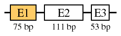
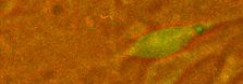
For the purposes of submission, we distinguish two types of digital images: presentation graphics (drawings, charts, diagrams, schematics) created de novo on the computer and bitmap graphics (gels, photographs, micrographs). Examples of these types of images are shown below:
Acceptable presentation formats are Postscript (.ps), Encapsulated Postscript (.eps), and Adobe Illustrator (.il7, .il8, or .il9). When preparing diagrams and charts, remember that your artwork will mostly be viewed on computer monitors; many readers will have difficulty with images wider than 500 pixels (screen dots).
Bitmaps should be submitted without compression and at a resolution of at least 144 dots per inch (if the data were collected to that resolution). Bitmap figures should be submitted without markings (arrows, labels, etc.). A lower resolution image should accompany the figure showing how the figure should be marked or labelled. Acceptable bitmap formats are TIFF (uncompressed) and Photoshop. For everyone's convenience, it is strongly recommended that you crop images to eliminate extraneous white space. If you submit images in Photoshop format, you may create additional layers to mark the images (do not flatten the images). When preparing bitmaps, remember that they will mostly be viewed on computer monitors; many readers will have difficulty with images wider than 500 pixels (screen dots).
Animation and Video (movies) should be submitted as a series of frames (TIFF). If you submit a quicktime movie, we strongly recommend that you keep the frames because editing compressed movies can cause severe degradation of quality. If you submit a series of frames, they should be numbered sequentially and all be of the same size. Each frame will be assumed to get equal display time unless otherwise noted.
Movies need to tread the line between being small enough in size for readers to download in a reasonable time and showing enough detail to make the movie a useful part of the report. When submitting a movie, you should also include an appropriate frame or set of frames to display to readers unable to view movies (these frames will also be used in the PDF version of articles).
Table 2 summarizes the formats that may be used for submitting figures digitally. For other types of figures (including sound and other data formats), the Editors should be consulted before preparing the figure.
Table 2. Acceptable formats for figures
|
Figure Type |
Acceptable Formats |
|
Presentation |
Illustrator (.il7, .il8, or .il9), Postscript (.ps), or Encapsulated Postscript (.eps) |
|
Bitmap |
TIFF (.tif) or Photoshop (.psd) |
|
Animation/Video |
TIFF (.tif) frames |
|
Other |
Consult the Editors before preparing other types of figures |
To reduce the size of graphic files, they may be compressed using Stuffit (.sit), Zip (.zip), or gzip (.gz). Please add the appropriate suffix to the file's name when you compress it.
Hypertext links
Molecular Vision does not consider hypertext links that point to locations external to the journal to be part of an article. During the galley process, the Editors will automatically add hypertext links to all GenBank accession numbers and to all references listed in PubMed. The Editors welcome the inclusion of other hypertext links with submissions. Hypertext links will be maintained in published articles, but if the target of a link disappears, the link will be removed.
When choosing hypertext links to submit, keep in mind that many web locations have a short lifetime by design. It is usually better to submit a link to a company that supplies a product described in the Methods section, than a link to the product itself. Since the hypertext link is not part of the manuscript, it can not serve to answer questions that should be covered in the text.
When mentioning gene or protein sequences, the Editors ask that they be named with GenBank numbers whenever possible. The Editors are committed to maintaining links to GenBank accession numbers as long as NCBI continues to make them available on the internet.
Sending your Submission
Files may be emailed to <molvis@emory.edu> using one of the following methods:
- For text files, copy the contents of the file into the body of an email message (one file per email message)
- Send files as MIME attachments to an email message (any number of files)
- Contact the Editors about submitting large files by FTP
3.5" disks (Mac, DOS/Win format) and/or "Zip" cartridges (Mac, DOS/Win, OS/2 format) may be mailed to:
Molecular Vision
c/o Jeffrey H. Boatright, Ph.D.
Lab B-5500, Emory Eye Center
1327 Clifton Road, N.E.
Atlanta, GA 30322
For information on the review and publishing process, see Publishing with Molecular Vision.
Editorial Board
|
Ruben Adler
Johns Hopkins University
Baltimore, MD, USA
Patricia D'Amore
Schepens Eye Research Institute
& Harvard Medical School
Boston, MA, USA
Cristina Arruti
Universidad de la Republica
Montevideo, Uruguay
Bronwyn J. Bateman
University of Colorado
Aurora, CO, USA
Steven L. Bernstein
University of Maryland
Baltimore, MD, USA
Joseph C. Besharse
Medical College of Wisconsin
Milwaukee, WI, USA
Suraj Bhat
Jules Stein Eye Institute, UCLA
Los Angeles, CA, USA
Diane Borst
Uniformed Service University
of Health Science
Bethesda, MD, USA
Greg Cahill
University of Houston
Houston, TX, USA
Deborah Carper
National Eye Institute, NIH
Bethesda, MD, USA
Gerald J. Chader
The Foundation Fighting Blindness
Hunt Valley, MD, USA
Ana B. Chepelinsky
National Eye Institute, NIH
Bethesda, MD, USA
Yves Courtois
Development, Aging and Pathology
of the Retina, INSERM U450
Paris, France
James Crabbe
University of Reading
Whiteknights, Reading, UK
Cheryl Craft
Keck School of Medicine, USC
Los Angeles, CA, USA
Rosalie Crouch
Medical University of South Carolina
Charleston, SC, USA
Stephen P. Daiger
University of Texas Health Science Center
Houston, TX, USA
Debora B. Farber
Jules Stein Eye Institute, UCLA
Los Angeles, CA , USA
Donald A. Fox
University of Houston
Houston, TX, USA
James L. Funderburgh
University of Pittsburgh
Pittsburgh, PA, USA
Federico Gonzalez-Fernandez
University at Buffalo,
The State University of New York
Buffalo, NY, USA
Carla Green
University of Virginia
Charlottesville, VA, USA
William Hauswirth
University of Florida
Gainesville, FL, USA
James Fielding Hejtmancik
National Eye Institute, NIH
Bethesda, MD, USA
Leonard M. Hjelmeland
University of California, Davis
Davis, CA, USA
James Hurley
University of Washington
Seattle, WA, USA
P. Michael Iuvone
Emory University
Atlanta, GA, USA
Winston Kao
University of Cincinnati
Cincinnati, OH, USA |
Gordon K. Klintworth
Duke University Medical Center
Durham, NC, USA
Nicolette H. Lubsen
University of Nijmegen
Toernooiveld Nijmegen, Netherlands
Mary Lyon
Medical Research Council
Harwell, UK
Stephen C. Massey
University of Texas Medical School
Houston, TX, USA
Peter Mathers
West Virginia University School of Medicine
Morgantown, WV, USA
Robert S. Molday
University of British Columbia
Vancouver, British Columbia, Canada
Mark Petrash
Washington University
St. Louis, MO, USA
Joram Piatigorsky
National Eye Institute, NIH
Bethesda, MD, USA
Mary E. Pierce
SUNY Upstate Medical University
Syracuse, NY, USA
Gadiparthi N. Rao
University of Tennessee
Memphis, TN, USA
T. Michael Redmond
National Eye Institute, NIH
Bethesda, MD, USA
Harris Ripps
University of Illinois at Chicago
Chicago, IL, USA
Lawrence Rizzolo
Yale University
New Haven, CT, USA
John (Jack) C. Saari
University of Washington
Seattle, WA, USA
Gail M. Seigel
University at Buffalo,
The State University of New York
Buffalo, NY, USA
Sue Semple-Rowland
University of Florida McKnight Brain Institute
Gainesville, FL, USA
Christine Slingsby
Birkbeck College
London, UK
Kent W. Small
Jules Stein Eye Institute, UCLA
Los Angeles, CA, USA
Deborah Stenkamp
University of Idaho
Moscow, ID, USA
Stephen Sugrue
University of Florida
Gainesville, FL, USA
Anand Swaroop
University of Michigan
Ann Arbor, MI, USA
Larry Takemoto
Kansas State University
Manhattan, KS, USA
Daniel Tranchina
New York University
New York, NY, USA
Robert Williams
University of Tennessee
Memphis, TN, USA
Graeme Wistow
National Eye Institute, NIH
Bethesda, MD, USA
Paul Wong
University of Alberta
Edmonton, Alberta, Canada
Donald Zack
Johns Hopkins Medical School
Baltimore, MD, USA
Peggy Zelenka
National Eye Institute, NIH
Bethesda, MD, USA |
|